High-Frequency QRS Analysis: From the Research Lab to the Clinical Market |
|---|
By Guy Amit, PhD., Biological Signal Processing Ltd., Tel-Aviv, Israel |
Ischemic heart disease (IHD) is a major healthcare problem worldwide. In the US alone, there are 1 7 . 6 million IHD patients, and the estimated annual incidence of new and recurrent myocardial infarction is estimated to be 935,000 (1). ECG retains its central role as the first-line diagnostic modality for ischemic heart disease workup in both chronic and acute presentations. Electrocardiographic detection of myocardial ischemia is typically based upon identification of repolarization abnormalities, manifested as changes in the ST-T segment. However, ischemia also induces changes during the depolarization phase, which can be detected and quantified by analyzing the high-frequency components of the QRS complex (HFQRS). HFQRS components are very low in amplitude and are typically filtered out by conventional ECG devices. The HFQRS signal originates from the fragmentation of the electrical activation wavefront in the myocardium, caused by the branching nature of the conduction system. In ischemic regions of the myocardium, local slowing of conduction velocity reduces the wavefront fragmentation, causing changes in the intensity and morphology of HFQRS signals (2). Over the last 30 years, HFQRS have been extensively studied in computer simulations, animal models, humans during coronary balloon occlusion and patients undergoing exercise test (2-7). Analysis of HFQRS has been reported to be more sensitive and specific than conventional ECG interpretation in detecting exercise-induced ischemia (5-7). Recently, Recently, BSP – Biological Signal Processing Ltd. (Tel Aviv, Israel) has introduced the HyperQ™ EX300 stress system, which provides automated HFQRS analysis in a commercially-available exercise ECG system. The system's technology is based upon sophisticated signal processing algorithms that perform accurate QRS alignment and averaging, band-pass filtering in the frequency range of 150 to 250 Hz, and computing of the root-mean-square intensity of the HFQRS signal. The trend of HFQRS intensity in 12 leads throughout the exercise test is then examined, and the relative intensity reduction is used as an index of ischemia (Figure 1).
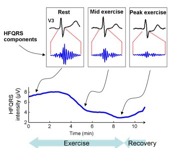
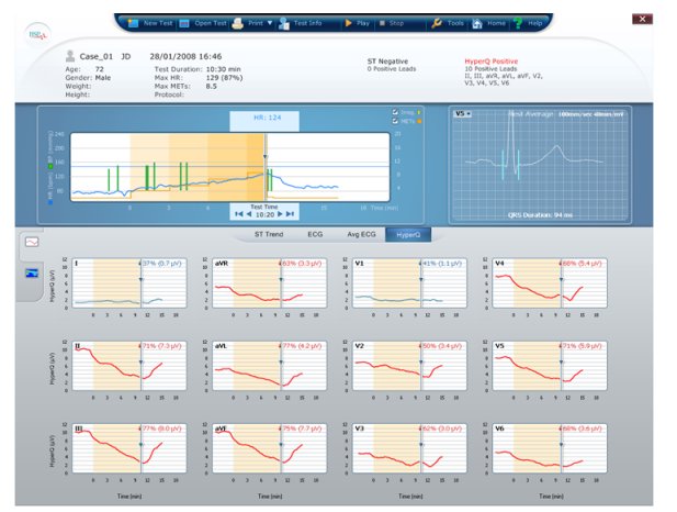
FIGURE 1:
Figure 1: Example of conventional ECG and HFQRS obtained in an ischemic patient (top), and the HFQRS analysis provided by the HyperQ EX-300 system (bottom). Note the marked reduction in HFQRS intensity with exercise. (click to enlarge)
The HyperQ index of ischemia was studied in 996 patients (age 58.9±10.2 years, 670 men), referred for exercise nuclear scan, which was used as the gold standard of ischemia. HyperQ response correlated with severity of ischemia, and was more sensitive (69% vs. 39%, P<0.005) and more specific (86% vs. 82%, P<0.05) than ST segment analysis. Multivariate logistic regression indicated considerable incremental diagnostic value of HyperQ over pre-test likelihood of CAD and exercise parameters (7).
Ongoing research efforts are aimed to establish HFQRS analysis as a routine adjunct to workup of IHD, and to develop new clinical applications of the technology. Of particular interest are the utilization of HFQRS for early detection of acute myocardial ischemia in patients presenting with chest pain, continuous monitoring of patients in risk of transient ischemic episodes and identification of ischemia from intra-cardiac pacing leads.
References
- Lloyd-Jones D et al. Heart Disease and Stroke Statistics--2010 Update. A Report From the American Heart Association. Circulation 2010; 121:e46-e215.
- Abboud S et al.. Simulation of high-resolution QRS complex using a ventricular model with a fractal conduction system. Effects of ischemia on high-frequency QRS potentials. Circ Res 1991;68:1751-1760.
- Abboud S. Subtle alterations in the high-frequency QRS potentials during myocardial ischemia in dogs. Comput Biomed Res 1987; 20(4):384-395.
- Abboud S et al. Detection of transient myocardial ischemia by computer analysis of standard and signal averaged high frequency ECG in patients undergoing percutaneus transluminal coronary angioplasty. Circulation 1987;76:585-96.
- Pettersson J et al. Changes in high-frequency QRS components are more sensitive than ST-segment deviation for detecting acute coronary artery occlusion. J Am Coll Cardiol 2000;36:1827-34
- Toledo E et al. Detection of Stress-Induced Myocardial Ischemia from the Depolarization Phase of the Cardiac Cycle - A Preliminary Study. J Electrocardiol 2009; 42:240-9
- Sharir T et al. Detection of Stress-Induced Myocardial Ischemia Using Analysis of Depolarization Abnormalities. J Am Coll Cardiol 2009;53:A297
Contact: guy@bspmedical.com