Acute Myocardial Infarction |
|---|
IDENTIFICATION : E-HOL-03-0160-001 |
Patients (N=93) with acute myocardial infarction were identifed based on clinical symptomatic: sudden chest pain (typically radiating to the left arm or left side of the neck), shortness of breath, nausea, vomiting, palpitations, sweating, and anxiety.The exclusion criteria for the the AMI patients were : patients in non-sinus rhythm, patients with major co-morbidity such as malignancy, severe hepatic , renal or cerebral disease, etc. The patients whom had prior CABG wer ealso excluded (but not the patient with history non-CABG coronary revascularization).The Electrocardiograms were acquired using three pseudo-orthogonal lead configuration (X, Y and Z). There is a initial resting supine period for a 20 minutes duration before starting the ambulatory recording.
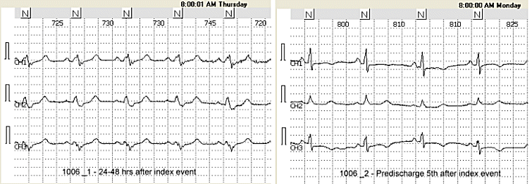
Sixty-seven patients with no prespecified conditions (age, gender, treatment) were enrolled with two Holter recordings performed: one between 24-48 after index event (ID_1) and the other predischarge between the 5th and 10th day after index event (ID_2). The remaining twenty six have one Holter recording (24-48 hrs after index event):
--- Patient ID 9019, 9023, 9067 9096 and 10001 had only second recording done, the other got only the first recording done.
All patients are suppose to be in sinus rhythm.
Exclusion criteria:
- the patients is in non-sinus rhythm (atrial flutter/fibrillation, pacemaker rhtyhm, AV block, sick sinus syndrome) during the first (24-48 hrs) Holter recording period,
- the patient has major comorbidity such as malignancy, severe haptic , renal or cerebral disease, etc.
- the patient had prior coronary artery bypass surgery (CABG) in the past; the patients with a history of non-CABG coronary revascularization (PTCA, stent, atherectomy) is eligible to be enrolled.
Basic descriptive statistics of the study population:
| MI location | Data | Nbfile single Holter | Follow-up Holter | All |
| Anterior | N | 9.0 | 11.0 | 20.0 |
| Age (yrs) | 66.7 | 66.1 | 66.4 | |
| BMI (kg/m2) | 25.8 | 26.7 | 26.3 | |
| EF (%) | 38.5 | 45.1 | 41.6 | |
| VT (n) | 0.0 | 1.0 | 1.0 | |
| syncope (n) | 1.0 | 0.0 | 1.0 | |
| Anterior lateral | N | 2.0 | 16.0 | 18.0 |
| Age (yrs) | 59.5 | 52.0 | 52.8 | |
| BMI (kg/m2) | 27.7 | 26.9 | 27.0 | |
| EF (%) | 48.5 | 55.5 | 54.5 | |
| VT (n) | 0.0 | 1.0 | 1.0 | |
| syncope (n) | 0.0 | 1.0 | 1.0 | |
| Inferior (posterior) | N | 6.0 | 21.0 | 27.0 |
| Age (yrs) | 59.0 | 54.4 | 55.4 | |
| BMI (kg/m2) | 24.5 | 27.1 | 26.5 | |
| EF (%) | 45.0 | 58.1 | 55.7 | |
| VT (n) | 0.0 | 1.0 | 1.0 | |
| syncope (n) | 0.0 | 0.0 | 0.0 | |
| Inferior lateral | N | 2.0 | 13.0 | 15.0 |
| Age (yrs) | 82.0 | 59.6 | 62.6 | |
| BMI (kg/m2) | 25.9 | 26.6 | 26.5 | |
| EF (%) | 54.5 | 50.1 | 50.8 | |
| VT (n) | 0.0 | 2.0 | 2.0 | |
| syncope (n) | 0.0 | 1.0 | 1.0 | |
| Unknown | N | 7.0 | 6.0 | 13.0 |
| Age (yrs) | 57.7 | 63.0 | 60.2 | |
| BMI (kg/m2) | 27.5 | 25.2 | 26.5 | |
| EF (%) | 51.0 | 55.0 | 52.0 | |
| VT (n) | 1.0 | 0.0 | 1.0 | |
| syncope (n) | 1.0 | 0.0 | 1.0 | |
| Total N | 63.1 | 57.5 | 59.1 | |
| Total Age (yrs) | 26.0 | 67.0 | 93.0 | |
| Total BMI (kg/m2) | 26.1 | 26.7 | 26.6 | |
| Total EF (%) | 44.6 | 53.8 | 51.2 | |
| Total VT (n) | 1.0 | 5.0 | 6.0 | |
| Total syncope (n) | 2.0 | 2.0 | 4.0 |
Total Clinical Information available for the database:
| ID | ID = 'Study Identifier' | ||
| nbFile | Number of files for this ID (1 or 2) | ||
| BETA_BLK | BETA_BLK = Taking Beta-Blockers(0/1) | ||
| DIGOXIN | DIGOXIN = Taking Beta-Blockers(0/1) | ||
| DIURETIC | DIURETIC = Taking Diuretics(0/1) | ||
| ACEINHIB | ACEINHIB = Taking Ace-Inhibitors(0/1) | ||
| ANTIARRH | ANTIARRH = Taking Anti-Arrhythmics(0/1) | ||
| FIBRINLY | Fibrinolysis (Y=1/N=0) | ||
| RACE | RACE = 'Race(White/Black/Asian/Other)' | ||
| HEIGHT | HEIGHT = 'Height (cm)' | ||
| WEIGHT | WEIGHT = 'Weight (Kg)' | ||
| BP_SYST | Systolic Blood pressure | ||
| BP_DIAST | Diastolic blood pressure | ||
| SMOKING | smoking (y=1/n=0) | ||
| HYPERTEN | HYPERTEN = 'Hypertension treated w/ Meds' | ||
| DIABETES | DIABETES = 'Diabetes Mellitus (0-no/1-w/insulin/2-w/o insulin)' | ||
| VT | VT = 'Ventricular Tachycardia' | ||
| SYNCOPE | SYNCOPE = 'Full Syncope(0/1)'; | ||
| PRIOR_MI | Date of Prior MI (mm/dd/yyyy) | ||
| AP | AP = 'Severity of Angina Pectoris' | ||
| CHF | CHF = 'Severity of CHF Treatment' | ||
| CPK | CPK = 'Peak CPK Level(I.U.)' | ||
| MI_LOCA | MI_LOC = 'Location of Infarction based on ECG' | ||
| 1 = 'Anterior' | |||
| 2 = 'Inferior(posterior)' | |||
| 3 = 'Lateral' | |||
| 4 = 'Ant-Lat' | |||
| 5 = 'Inf=Lat' | |||
| 9 = 'Unknown' | |||
| THROMBO | THROMBO = 'Thrombolytic Therapy' | ||
| PUL_CONG | PUL_CONG = 'Pulmonary Congestion(0/1)' | ||
| AP_3DAYS | AP_3DAYS = 'Angina Pectoris 1-3 days prior to discharge' | ||
| ST_DEP | ST_DEP = 'Transient ST segment Depression(0/1)' | ||
| VA | VA = 'Ventricular Arrhythmias' | ||
| 0 = 'NO VPBS' | |||
| 1 = 'VPBs' | |||
| 2 = 'NSVT(3beats/30secs)' | |||
| 3 = 'SVT(>30sec)' | |||
| 4 = 'Torsade de Pointes' | |||
| 5 = 'VF' | |||
| 6 = 'Other' | |||
| 9 = 'Unknown'; | |||
| LVDS | LV systolic dimension | ||
| LVDD | LV diastolic dimensions | ||
| REG_DATE | RECORDINGDATE = 'Date of the first recording' | ||
| AGE | AGE = 'Age' | ||
| GENDER | SEX = 'Sex(M/F)' | ||
| BMI | BMI = 'Body Mass Index' | ||
| EF | EF = 'Ejection Fraction via Angiogram/Echocardiogram' | ||
| WMA | WMA = 'Wall Motion Abnormality via Echo/Angiogram' | ||
ECG Characteristics:
- Number of leads : 3 (X, Y Z leads, pseudo orthogonal configuration)
- Sampling Frequency : 200Hz
- Amplitude Resolution: 10 uV
- Example of 10-min ECG and associated annotation files are availble for download: example.ecg and example.ann
- The format of these files is described in here.